Working Time
- Timings: 10:00 am to 1:00 pm – 5:00 pm to 7:00 pm
(Monday-Saturday)
Retinal Detachment Surgery

What is a retinal detachment?
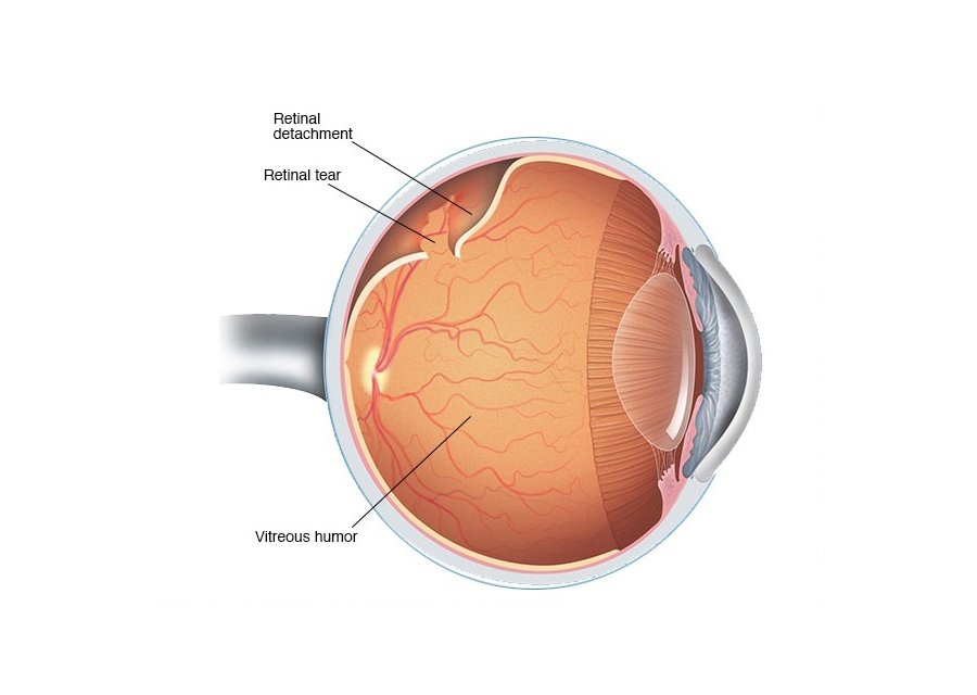
A retinal detachment is an eye condition involving separation of the retina from its attachments to the underlying tissue within the eye. Most retinal detachments are a result of a retinal break, hole, or tear. A retinal detachment of this type is known as a rhegmatogenous retinal detachment. Most retinal breaks, holes, or tears are not a result of injury. The majority of retinal breaks, holes, or tears are spontaneous, result when the vitreous gel pulls loose or separates from its attachment to the retina, usually in the peripheral parts of the retina.
How Is a Detached Retina Diagnosed?
Your ophthalmologist will put drops in your eye to dilate (widen) the pupil. Then they will look through a special lens to check your retina for any changes.
How is a retinal detachment repair performed?
There are several types of surgery to repair a detached retina. A simple tear in the retina can be treated with freezing, called cryotherapy, or a laser procedure. Different types of retinal detachment require different kinds of surgery and different levels of anesthesia. The type of procedure your doctor preforms will depend on the severity of retinal detachment. A vitrectomy surgery is done for serious retinal detachments. It may require partially removing the vitreous fluid inside the eye. Local anesthesia is used and the procedure is usually done in a surgical clinic.

